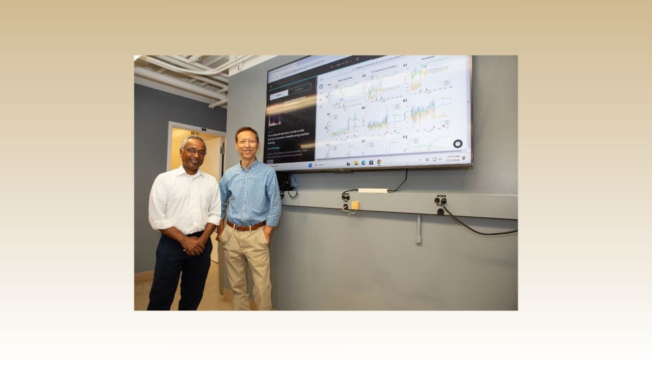
For decades, scientists have viewed seizures as moments when the brain’s neurons fire together in a synchronized storm of electrical activity.
New research from Purdue University reveals the brain’s story is far more complex.
By combining a novel laboratory model with advanced machine learning, researchers found that even during seizure-like activity, neurons organize into distinct functional subnetworks with their own unique firing patterns. The discovery offers new insight into how seizures develop and spread while demonstrating how artificial intelligence can help scientists uncover biological relationships that previously remained hidden.
The interdisciplinary research, led by Riyi Shi, the Mari Hulman George Endowed Professor of Applied Neuroscience in Purdue’s College of Veterinary Medicine and director of the Center for Paralysis Research, could one day improve seizure diagnosis, accelerate drug discovery and pave the way for more personalized treatments for epilepsy and other neurological disorders.
“Despite their prevalence, we still don’t fully understand the mechanisms behind seizure onset and progression,” Shi says. “Traditional methods tell us when seizure activity is happening, but they miss many of the subtle changes occurring within the neuronal network. By working with experts in machine learning, we were able to look much deeper into the data.”
The results challenged one of the long-held assumptions about how seizures unfold in the brain.
Looking beyond synchronized activity
For years, researchers have known that seizures involve highly synchronized electrical activity in the brain. The prevailing assumption has been that neurons largely behave as a single coordinated network during these events. Shi’s team discovered otherwise.
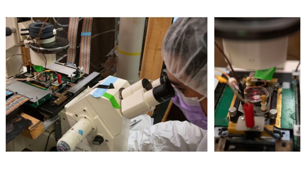
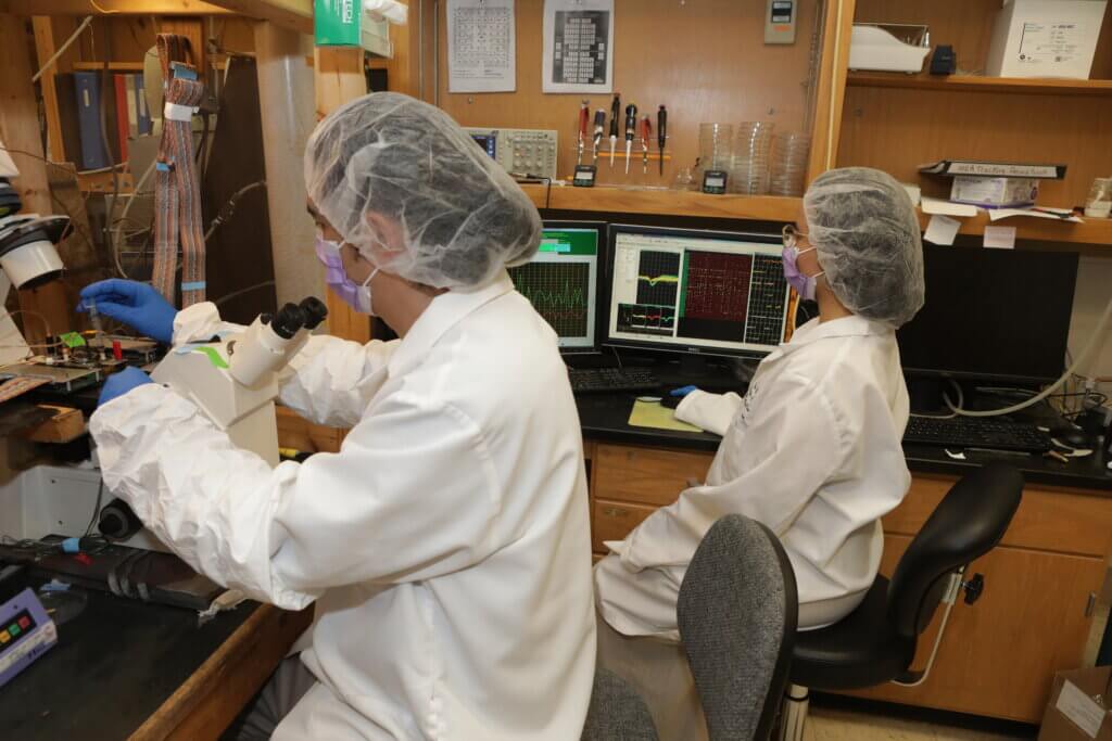
The researchers relied on an in vitro “seizure-on-a-chip” model — cultured animal brain cells grown on a microelectrode array that records electrical activity across neuronal networks — to generate detailed recordings of seizure-like events under carefully controlled laboratory conditions.
Because the neurons are grown in a controlled environment, researchers can observe how brain cells communicate without many of the variables present in living animals or people. The model allows the team to repeatedly study the same types of seizure-like activity while collecting detailed electrical recordings that would be difficult to isolate in more complex systems. Those recordings provide an ideal testing ground for evaluating new analytical approaches.
The recordings captured enormous amounts of information, far more than traditional analytical methods could fully interpret. Rather than relying on conventional measurements of synchronization alone, Shi partnered with faculty in Purdue’s College of Science, including Ananth Grama, the Samuel D. Conte Distinguished Professor of Computer Science and director of Purdue’s Institute for Physical AI, to apply machine learning techniques capable of recognizing complex relationships within the data.
To analyze those recordings, Grama’s team trained machine learning models on the university’s Gautschi high-performance computing cluster. The models identified distinct groups of neurons that consistently fired together over time, even when those neurons were physically distant.
“We uncovered subnetworks of neurons with distinct firing behaviors that would have been invisible to traditional analysis,” Grama says. “This represents a shift from viewing AI as a downstream analysis tool to AI as the method that reveals the underlying biophysical phenomenon itself.”
Collaborators included Aniket Bera, associate professor of computer science, and doctoral student researchers Shatha Mufti and Jhon Martinez (Shi Lab), and Shourya Verma (Grama Lab) and Prerit Gupta (Bera Lab).
Published in the Journal of Neurophysiology, the study demonstrates how combining experimental neuroscience with machine learning can reveal biological relationships that conventional analytical methods often overlook. The AI models also distinguished seizure activity from normal brain activity with greater sensitivity than traditional analytical approaches.
“We found that not all neurons behave the same way during seizure activity,” Shi says. “These hidden subnetworks suggest that functional connections between neurons — not simply where they are located — play an important role in how seizures develop.”
Building better tools for diagnosis and treatment
While the research was conducted using an in vitro model, the findings could have far-reaching implications.
Identifying these functional neuronal networks gives researchers new biological markers to investigate as they develop better diagnostic tools and evaluate potential therapies. The same platform could also accelerate drug screening by allowing scientists to measure how candidate treatments affect specific neuronal networks rather than seizure activity as a whole.
Better biological markers could also improve how researchers evaluate potential therapies. Instead of relying solely on whether seizure activity increases or decreases, scientists may eventually be able to measure how individual neuronal subnetworks respond to experimental drugs. That level of precision could help researchers identify promising therapies earlier in the development process while improving their understanding of how different treatments affect the brain.
Although the study focused on seizure-like activity, the analytical framework has applications beyond epilepsy. Similar approaches could help researchers study neurological disorders associated with traumatic brain injury as well as conditions involving learning, memory and other complex brain functions.
The approach could eventually contribute to more personalized care for patients living with epilepsy and other neurological disorders. Today, seizures often remain difficult to predict before symptoms appear. In the future, Shi envisions that AI-powered monitoring systems will be capable of recognizing subtle changes in brain activity, potentially alerting patients or clinicians before a seizure occurs.
“The goal is not simply to detect seizures,” Shi says. “It’s to better understand the underlying biology well enough that we can improve diagnosis, treatment and ultimately patients’ quality of life.”
A model for interdisciplinary discovery
The project also showcases Purdue’s growing strength in bringing together researchers from different disciplines to tackle problems that no single field could solve alone. Shi’s laboratory generated the experimental data, while Grama’s group developed computational approaches capable of extracting biological signals that would have been difficult to detect through conventional analysis.
The collaboration also illustrates how AI is changing the scientific process itself. Rather than serving only as a tool for analyzing completed experiments, machine learning is becoming a partner in discovery by identifying meaningful biological signals that scientists can then investigate further.
“This project demonstrates the power of AI models to extract biologically meaningful, potentially diagnostic signals from data that’s otherwise difficult to interpret,” Grama says. “Purdue’s culture of collaboration and its investment in physical AI create an environment where these kinds of discoveries can happen.”
The research also aligns with the university’s One Health initiative by combining veterinary neuroscience, computer science and advanced computing to generate discoveries with potential relevance to both animal and human neurological health. According to Shi, that interdisciplinary approach is essential for tackling some of the brain’s most complex challenges.
“The future of neuroscience will require biology, medicine, engineering and artificial intelligence to work together,” he says. “When you bring those disciplines together, you can begin to answer questions that have challenged researchers for many years.”