
USDA-NIFA award funds project to decode the earliest “conversation” of pregnancy and reduce embryonic loss
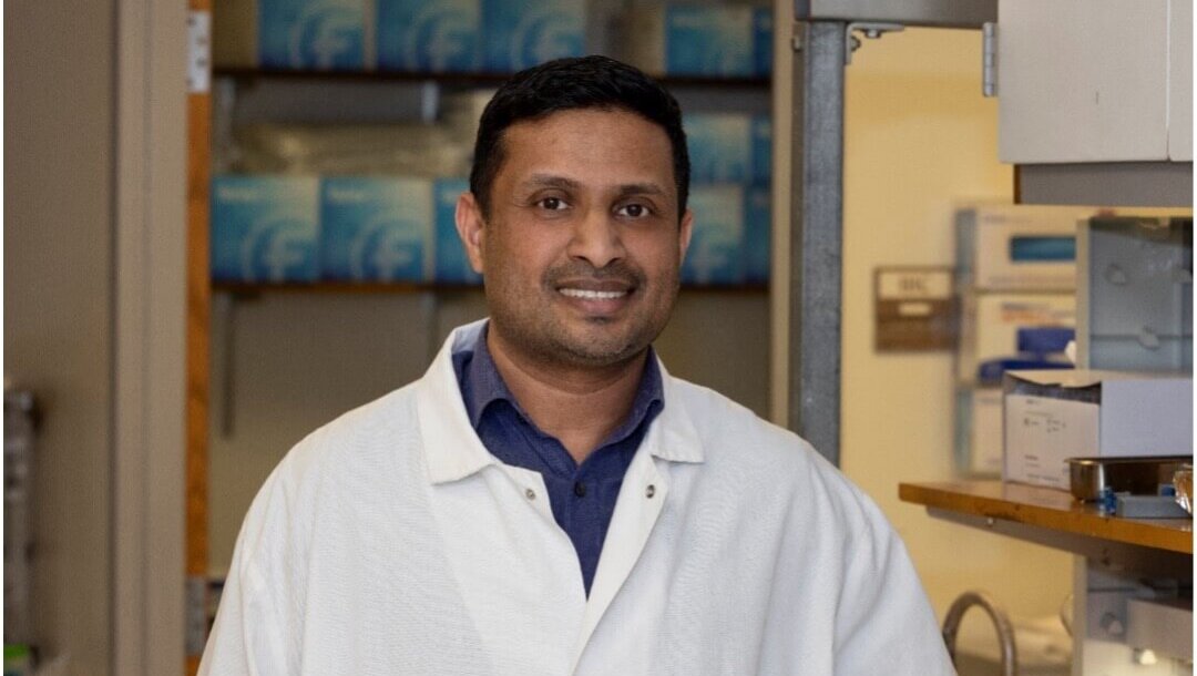
A four-year, $650,000 New Investigator Award from the USDA National Institute of Food and Agriculture (USDA-NIFA) will support research led by Dr. Viju V. Pillai, a faculty member in Purdue Veterinary Medicine’s Department of Comparative Pathobiology and pathologist at the Animal Disease Diagnostic Laboratory (ADDL), aimed at solving a persistent and costly mystery in cattle production. Dr. Pillai’s team wants to answer the question of why so many pregnancies fail before they are even recognized? The project will focus on the earliest stages of fetal–maternal communication and on a little-understood family of proteins called trophoblast Kunitz domain proteins (TKDPs), whose functions in pregnancy remain largely unknown.
“Pregnancy does not begin with a visible sign—it begins with a signal,” said Dr. Pillai, assistant professor of anatomic pathology and the project’s lead investigator at Purdue. “Within the first week, the embryo must effectively ‘introduce itself’ to the mother. If that molecular dialogue falters, the pregnancy can quietly end before it truly begins.”
Early embryonic loss is a major challenge in modern cattle systems. Dr. Pillai says even though advances in genetics and production have improved milk and meat yield, reproductive efficiency has not kept pace. The first few weeks of pregnancy are especially vulnerable, and failures during this window can lead to substantial economic losses for producers through missed breeding cycles, extended calving intervals, and reduced lifetime productivity.
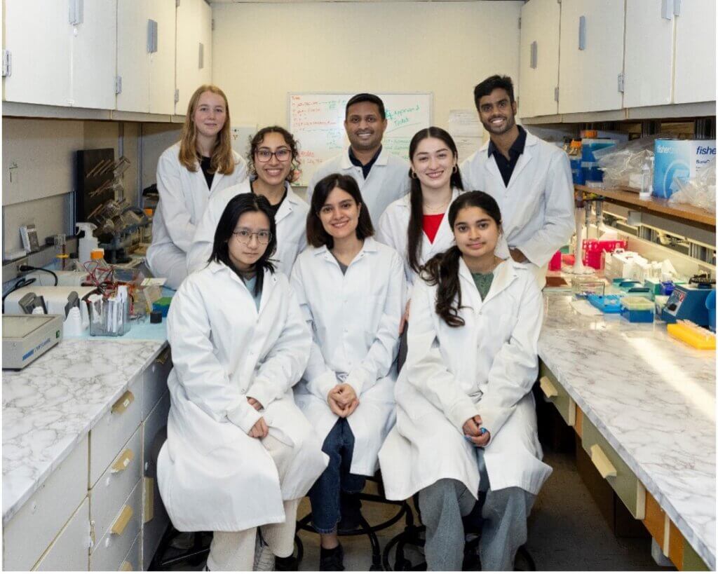
To address this challenge, Dr. Pillai’s Laboratory of Developmental and Stem Cell Biology is developing and deploying experimental models that recreate key features of early pregnancy outside the animal, creating an accessible “window” into events that are otherwise difficult to observe. Central to this work are trophoblast cells, the specialized cells that build the placenta and serve as an essential interface between the embryo and the uterus. By growing these cells under carefully controlled conditions that mimic the uterine environment, the team has established platforms in which trophoblast cells not only survive but also differentiate into placenta-forming cell types that resemble those seen in early gestation.
Dr. Pillai says the newly funded research builds on preliminary findings using organoid models called “trophocysts” – tiny, embryo-like spheres composed entirely of trophoblast cells. These structures resemble aspects of an early conceptus but lack the fetal component that would develop into a calf. In initial proof-of-concept studies, a subset of recipients receiving these trophocysts showed biological responses consistent with early pregnancy recognition, suggesting that trophoblast-derived signals may play a direct role in triggering maternal adaptation.
With the USDA-NIFA support, the team will advance this work using CRISPR-based gene editing to precisely alter candidate genes in trophoblast cells and then generate engineered trophocysts to test how specific molecular changes influence the maternal response. A major emphasis will be placed on TKDPs, a set of trophoblast-expressed proteins that are strongly implicated in ruminant pregnancy biology but remain functionally enigmatic. By selectively modifying TKDP-related pathways and measuring downstream responses, the project aims to define whether these proteins contribute to successful pregnancy establishment and maintenance.
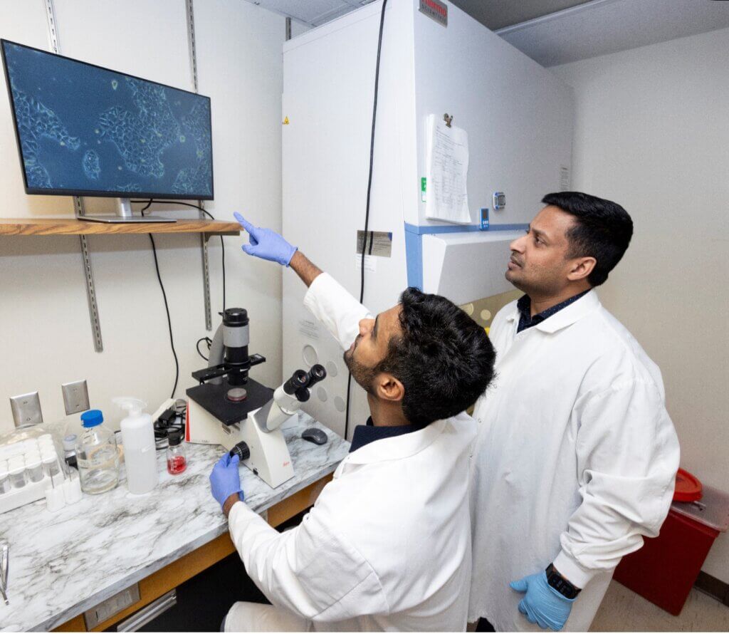
The work also will extend beyond the earliest “hello” of pregnancy. After maternal recognition occurs, pregnancy is sustained through specialized structures, including placentomes, where uterine and placental tissues interlock to exchange nutrients and signals. Pillai’s laboratory has developed three-dimensional trophoblast culture systems that form villus-like projections – features that mirror the placenta’s exchange surface, and can be used to study how hormones, nutrients, and molecular cues traverse the maternal-fetal interface as gestation progresses.
The long-term goal is practical and translational: identify actionable biological mechanisms that can reduce reproductive losses and improve sustainability in livestock production. Beyond agriculture, the work also aligns with Purdue University’s One Health strategic initiative. Dr. Pillai explains that many principles of embryo-maternal communication are shared across mammals – the underlying “language” is conserved even when the species-specific details differ. Insights from cattle may therefore inform broader questions in reproductive biology, including mechanisms relevant to infertility and early pregnancy loss in people.
“On the surface, this is a story about cattle reproduction,” Dr. Pillai said. “But at its core, it is a story about how life begins, through a silent exchange long before a calf ‘moos’ or a baby cries. By decoding that earliest dialogue, we can support healthier herds, improve efficiency for producers, and deepen our understanding of pregnancy across mammals.”
