
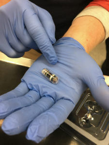
This pill, about the size of a large vitamin, is equipped with a light source and an array of photo sensors that captures detailed images of the GI tract in small animals. Capsule endoscopy has been around since the late 1990s for human patients, but only recently has been developed for the veterinary industry, and now is being used at PVM’s Veterinary Teaching Hospital.
One of the many challenges veterinarians face is that animals can’t tell them exactly what’s wrong. But now, with a new camera capsule available from ALICAM at the Purdue University Veterinary Teaching Hospital (VTH), Purdue veterinary specialists can gain valuable insights into their patients’ gastrointestinal health with no harm, no anesthesia, and no recovery time required. Capsule endoscopy has been around since the late 1990s for human patients, but has only recently been developed for the veterinary industry.
“It’s as simple as giving a dog a pill,” said Dr. Andrew Woolcock, assistant professor of small animal internal medicine in the College of Veterinary Medicine’s Department of Veterinary Clinical Sciences. “After you administer the endoscopy pill, the pill travels through the GI tract recording high-quality 360 degree images. We used to have to sedate the animal and do a full endoscopy to get imaging of the digestive tract. But one of the limits of that method, because the intestines are so long, is that there’s always some of the tract that is invisible to the traditional scope. With the camera pill we have the advantage of seeing the complete digestive tract, it’s less invasive for the patient, and it takes the guesswork out of GI diagnoses.”
Like endoscopy pills for humans, the veterinary version is about the size of a large vitamin. It uses an array of photo sensors and an internal light source to generate high-resolution images in patients, taking thousands of images with a miniature camera.
After the dog takes the pill, the owner must recover the pill and is provided with a pre-paid shipping envelope to send it back for interpretation. Board certified specialists analyze the images for abnormalities and design treatment options based on their findings. The pill is not the same as a biopsy, as it doesn’t provide microscopic images, but it might establish a definitive diagnoses, like ulcers, or it may indicate that other tests such as biopsies need to be done.
“This is a great option for pet owners who are worried about invasive interventions, but want to investigate GI symptoms in their pets,” said Dr. Woolcock, who is board certified in small animal internal medicine. Symptoms such as weight loss, reduced appetite, food intolerance, vomiting, diarrhea, abdominal pain, unexplained anemia (low red blood cell count), and dark or bloody stools indicate the need for diagnostic imaging to help diagnose a dog’s specific medical condition.