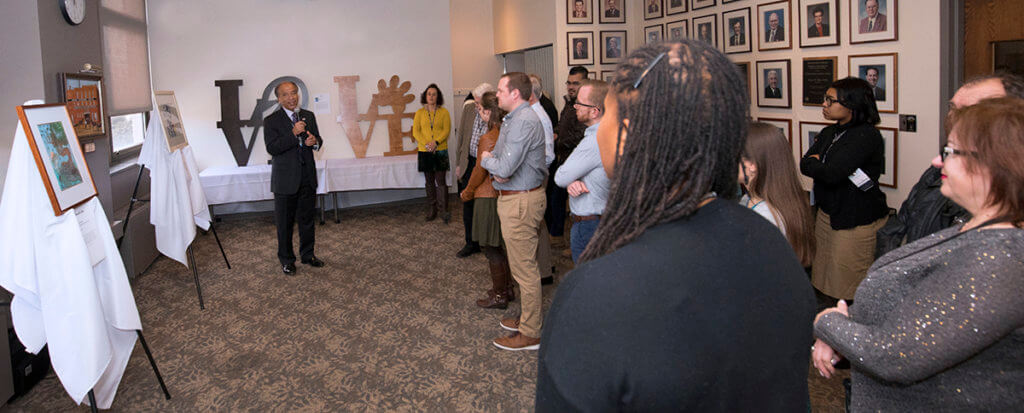
Purdue Veterinary Medicine faculty, staff, students, and guests gathered Friday afternoon, February 22, in the Alumni Faculty Lounge (Lynn 1192) for a special ceremony to dedicate five new works of art that will be displayed in the College of Veterinary Medicine. “This is always a day that I look forward to because I get to see the wonderful new creations of beautiful art that will be on the walls and in our building we hope for many, many years,” Dean Willie Reed said as he welcomed everyone to the event.
The Art in Lynn Hall program started in the spring of 2010, with a call for members of the Purdue Veterinary Medicine family to submit proposals. The initial set of three pieces was dedicated in 2011. “We started this with the idea of, ‘wouldn’t it be wonderful to have members of the PVM family contribute original art that we could display in our college?’… And so, with the help of Professor David Williams, who has really led this project from the beginning, it became a reality,” Dean Reed recalled. “I’m just always amazed at the talent that we have here amongst our students, our faculty, and our staff, and even some of the relatives of our family here in Lynn Hall.” Dean Reed then introduced each of the artists, who had the opportunity to describe their work.
The five new pieces were created by Dr. Tiffany Lyle, assistant professor of veterinary anatomic pathology in the Department of Comparative Pathobiology; Thane Boyce of the DVM Class of 2020; Emily Matson of the Veterinary Nursing Class of 2019; Joni Montgomery of the DVM Class of 2022; and James Woolcock, who is the father of Dr. Andrew Woolcock, assistant professor of small animal internal medicine.
The artists provided the following statements about their work:
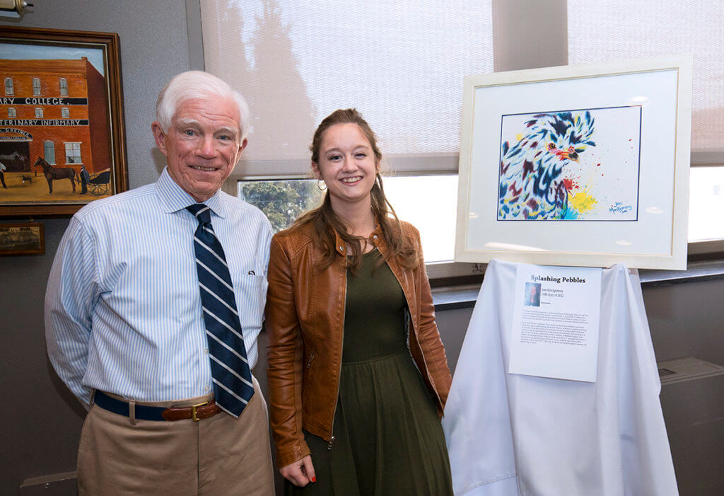
“Splashing Pebbles”, watercolor by Joni Montgomery, of the DVM Class of 2022
The field of poultry medicine is always growing and changing. Over the last few years, backyard poultry has grown exponentially in popularity. Chickens are playing a bigger role in the human-animal bond and there is a need for more veterinarians to be able to see and treat chickens as the pets they have become.
As an avid chicken hobbyist, I love seeing my birds’ personalities and colors shine through as I get to know them individually. I wanted to create a painting of a chicken that was full of color and personality to remind others that even chickens play an important role in the human-animal bond. This painting is of one of my own birds I raised from a chick. Pebbles, a Mottled Houdan, won ribbons at multiple poultry shows around the state and I enjoyed having her in my life until she passed away of natural causes. Pebbles was a funny little character and so full of life – I felt she would be the perfect model to portray the goal of my artwork.
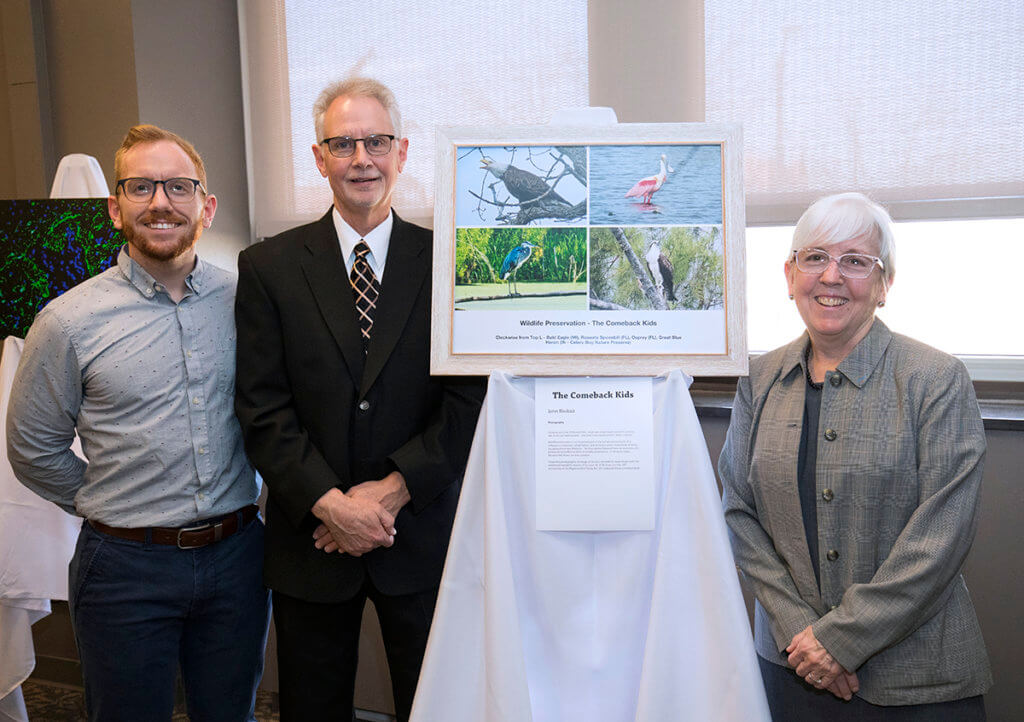
“The Comeback Kids”, photography by James Woolcock, father of Dr. Andrew Woolcock, assistant professor of small animal internal medicine
Growing up in the 1950s and ‘60s, I never saw a bald eagle except in pictures. Yet, it was our national bird. I was told it was nearly extinct. What a shame!
Wildlife preservation is an important part of the human-animal bond. It is reflected in treatment, rehabilitation, and advocacy across many fields of study, including Veterinary Medicine. The four species featured here are examples of a protracted and effective ethic of wildlife preservation. It still exists today because the threats are ever-present. I hope this photographic montage serves as a reminder to never forget both the sordid and wonderful lessons of our past. As 2018 [marked] the 100th anniversary of the Migratory Bird Treaty Act, let’s celebrate these comeback kids!
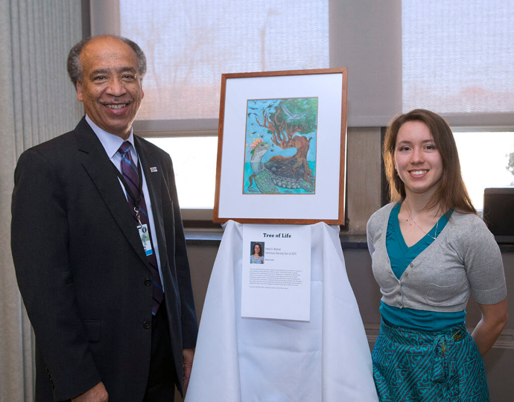
“Tree of Life”, mixed media by Emily A. Matson, of the Veterinary Nursing Class of 2019
The tree of life built from life is meant to represent the connectivity of nature to one another and the diversity of life on earth. All life from insect to great elephant, from invertebrate to avian to amphibian to reptilian to mammalian, even to the mythical as the tree grows on the great world turtle of Native American creation myth. Connectivity is shown with each creature building the trunk, branches, and leaves, displaying that not one life is greater than the other, the various animals are all connected, twisting and folding upon one another to create the grain, branches, foliage, and roots of the tree in order to survive.
Can you identify all the creatures? Can you find the human?
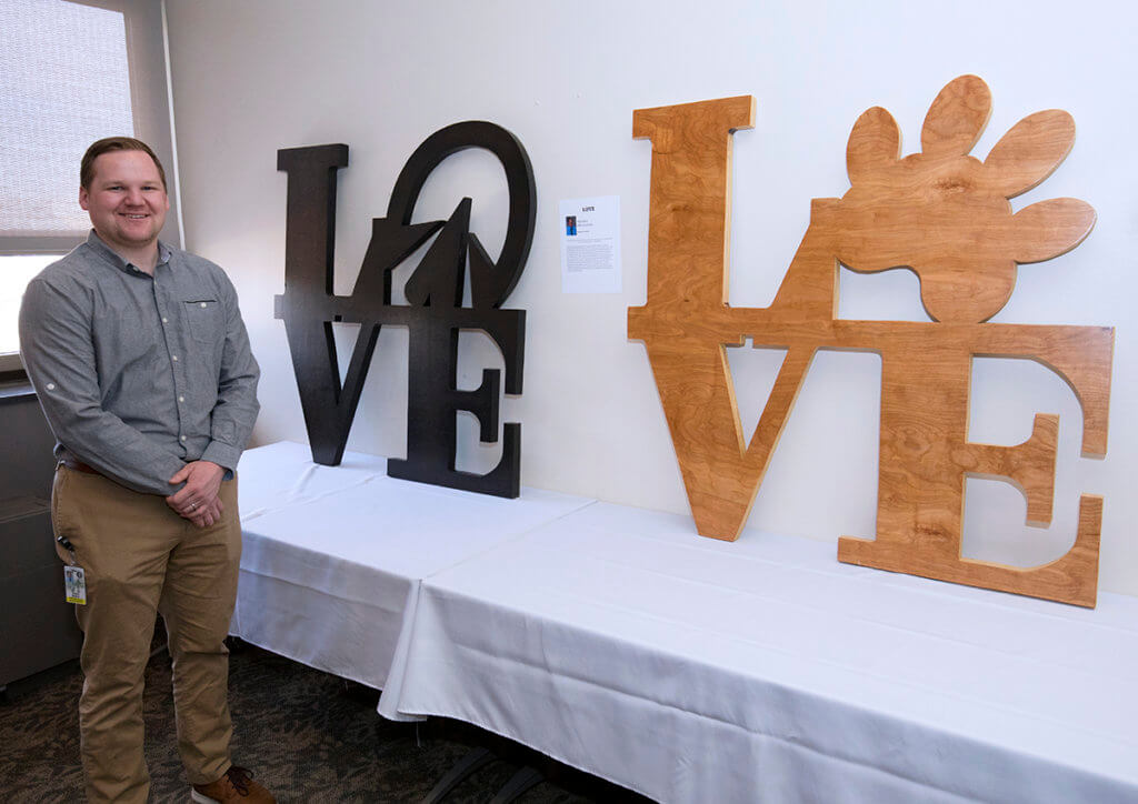
“LOVE”, wooden sculptures by Thane Boyce, of the DVM Class of 2020
These sculptures were inspired by the work of Robert Indiana, a native of Indiana who designed the famous LOVE sculptures seen around the world. The bond between humans and animals can be one of the strongest expressions of love we experience. There are countless stories of the sacrifices that humans and animals make for each other that demonstrate just how strong that love and loyalty can be. These sculptures are dedicated to all of those expressions of love. The sculptures are made from wood to show how natural that bond is. Small defects in the wood and creation process were left to illustrate that love isn’t perfect, but that in the end it creates something beautiful. The dog paw print and horse hoofprint were chosen to emphasize that our love isn’t limited to one species but includes all animals from the small and large animal world. The black and gold stains were chosen to highlight Purdue’s dedication to the animals that we love.
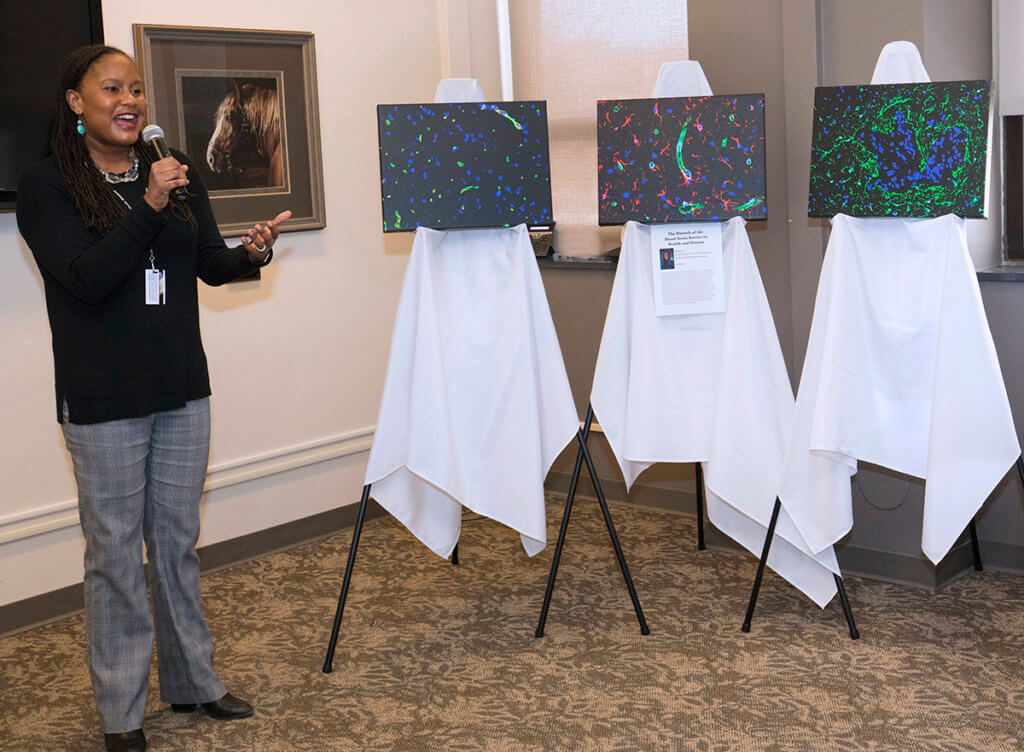
“The Marvels of the Blood-Brain Barrier in Health and Disease”, photography by Dr. Tiffany Lyle, assistant professor of veterinary pathology in the Department of Comparative Pathobiology
As a veterinary pathologist and Principal Investigator in the Comparative Blood-Brain Barrier Research Laboratory, I regularly reflect on the high impact images that reflect veterinary medicine in the diagnostic and research settings. This art installation represents images of the blood-brain barrier, the tightest and most effective barrier in the body, acquired from our laboratory. These fascinating images of the blood-brain barrier in an experimental mouse model of lung cancer were acquired using a fluorescence microscope. Panel A demonstrates the normal distribution of capillaries, highlighted in yellow, throughout a normal section of brain. In Panel B, as tumor cells begin to disseminate through the neuroparenchyma, the dilated vasculature (green) is infiltrated by astrocytes (red). Lastly, in Panel C, as a solid tumor forms in the brain there is a marked neuroinflammatory or neuroprotective response composed primarily of astrocytes (green) in the periphery of the tumor (blue). Altogether, Panels A-C demonstrate the changes in the blood-brain barrier in a disease state. The goal of this display is to highlight the cutting-edge research in our college, and encourage onlookers to take a moment to reflect on the depth and breadth of research and animal models in veterinary medicine.
