BMS Professor Among Team of Purdue Scientists Using Doppler to Peer Inside Cells in Effort to Improve Infection Diagnoses and Treatments

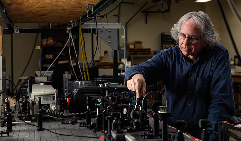
Professor David Nolte using Doppler radar.
Dr. David Nolte works with the Doppler apparatus as part of a research team using Doppler to peer inside living cells and gain insight into intracellular activity, metabolism, and pathogenicity. (Purdue University photo/Rebecca McElhoe)

Doppler radar improves lives by peeking inside air masses to predict the weather. A Purdue University team that includes Dr. John Turek, professor of basic medical sciences, is using similar technology to look inside living cells, introducing a method to detect pathogens and treat infections in ways that scientists never have before.

In a new study, the team used Doppler to sneak a peek inside cells and track their metabolic activity in real time, without having to wait for cultures to grow. Using this ability, the researchers can test microbes found in food, water, and other environments to see if they are pathogens, or help them identify the right medicine to treat antibiotic-resistant bacteria.

Dr. David Nolte, Purdue’s Edward M. Purcell Distinguished Professor of Physics and Astronomy; Dr. Eduardo Ximenes, research scientist in the Department of Agricultural and Biological Engineering; Dr. Michael Ladisch, Distinguished Professor of Agricultural and Biological Engineering, and Dr. Turek adapted this technique from their previous study on cancer cells in a paper released this month in Communications Biology.

Using funding from the National Science Foundation as well as Purdue’s Discovery Park Big Idea Challenge, the team worked with immortalized cell lines — cells that will live forever unless you kill them. They exposed the cells to different known pathogens, in this case salmonella and E. coli. They then used the Doppler effect to spy out how the cells reacted. These living cells are called “sentinels,” and observing their reactions is called a “biodynamic assay.”

“First we did biodynamic imaging applied to cancer, and now we’re applying it to other kinds of cells,” Dr. Nolte said. “This research is unique. No one else is doing anything like it. That’s why it’s so intriguing.”

This strategy is broadly applicable when scientists have isolated an unknown microbe and want to know if it is pathogenic — harmful to living tissues — or not. Such cells may show up in the food supply, water sources or even in recently melted glaciers.
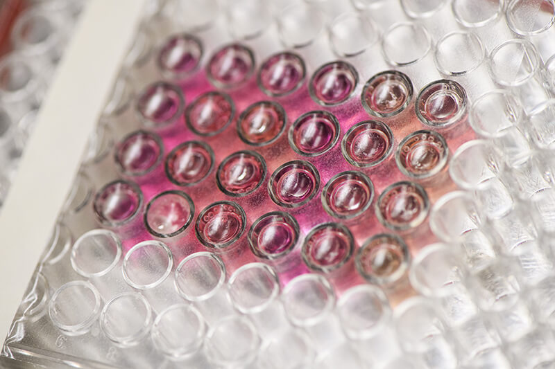
A multi-cell plate from Dr. David Nolte's lab.
The team isolated living immortalized cells in multi-well plates to study them with Doppler. (Purdue University photo/Rebecca McElhoe).

“This directly measures whether a cell is pathogenic,” Dr. Ladisch said. “If the cells are not pathogenic, the Doppler signal doesn’t change. If they are, the Doppler signal changes quite significantly. Then you can use other methods to identify what the pathogen is. This is a quick way to tell friend from foe.” 

Being able to quickly discern whether a cell is harmful is incredibly helpful in situations where people encounter a living unknown microorganism, allowing scientists to know what precautions to take. Once it is known that a microbe is harmful, they can begin established protocols that allow them to determine the specific identity of the cell and determine an effective antibiotic against the microorganism.

Another benefit is the ability to quickly and directly diagnose which bacteria respond to which antibiotics. Antibiotic resistance can be a devastating problem in hospitals and other environments where individuals with already compromised bodies and immune systems may be exposed to and infected by increasingly high amounts of antibiotic resistant bacteria. Sometimes this results in a potentially fatal condition called bacterial sepsis, or septicemia. This is different from the viral sepsis that has been discussed in connection with COVID-19, though the scientists say their next steps will include investigating viral sepsis.

Treating sepsis is challenging. Giving the patient broad-spectrum antibiotics, which sounds like a good idea, might not help and could make the situation worse for the next patient. Letting bacteria come into close contact with antibiotics that do not kill them only makes them more resistant to that antibiotic and more difficult to fight the next time.

Culturing the patient’s tissues and homing in on the correct antibiotic to use can take time the patient does not have, usually eight to 10 hours. This new biodynamic process allows scientists to put the patient’s bacterial samples in an array of tiny petri dishes containing the tissue sentinels and treat each sample with a different antibiotic. Using Doppler, they can quickly notice which bacterial samples have dramatic metabolic changes. The samples that do are the ones that have reacted to the antibiotic — the bacteria are dying, being defeated and beaten back by antibiotics.

“When we treat with antibiotics, the bacteria don’t have to multiply much before they start to affect the tissue sentinels,” Dr. Nolte explained. “There are still too few bacteria to see or to measure directly, but they start to affect how the tissues behaves, which we can detect with Doppler.”

In less than half the time a traditional culture and diagnosis takes, doctors could tell which antibiotic to administer, bolstering the patient’s chances for recovery. The researchers worked closely with the Purdue Research Foundation Office of Technology Commercialization to patent and license their technologies. They plan to further explore whether this method would work for tissue samples exposed to nonliving pathogenic cells or dried spores, and to test for and treat viral sepsis.

Click here to view a complete news release about the research.

Writer(s): Brittany Steff | pvmnews@purdue.edu

Recent Stories

Purdue Veterinary Medicine’s Connection with Wildlife Rescue in Guatemala Highlighted during International Education Week

Hidden amid the dense forests near the shores of Lake Petén Itzá, the ARCAS Wildlife Rescue Center is one of Central America’s most active sanctuaries for endangered and trafficked wildlife. In addition to their conservation efforts, ARCAS (Asociación de Rescate y Conservación de Vida Silvestre) offers unparalleled hands-on experience for veterinary and biology students from around the world. The Purdue University College of Veterinary Medicine has proudly collaborated with ARCAS for many years, and International Education Week this week (November 16-22) provided the perfect opportunity to showcase this invaluable educational partnership.

Immersive Mixed Reality Experience Helps Purdue Veterinary Students Learn Cardiac Physiology

First year Doctor of Veterinary Medicine students at the Purdue University College of Veterinary Medicine recently stepped into a new era of learning — an immersive Mixed Reality (MR) experience at Purdue’s Envision Center. This cutting-edge session allowed students to interact with a dynamic, 3D representation of the heart, deepening their understanding of cardiac physiology in ways traditional methods cannot match.

Feathered Fame: Purdue Veterinary Medicine Research Featured on Journal Cover

The Department of Veterinary Clinical Sciences (VCS) at Purdue University is proud to announce that a recent study from its anesthesiology team has been selected as the cover feature for Veterinary Sciences (MDPI), Volume 12, Issue 11. Chosen from among 82 articles, the publication highlights the College of Veterinary Medicine’s growing impact in avian clinical research.

“Paws Up” – brought to you by the PVM Wellness Committee

Time to shine a spotlight on some “paws-itively” amazing work by one of our newest Purdue Veterinary Medicine staff members, Jennifer Hartman, VCS Curriculum Technologist in the Department of Veterinary Clinical Sciences.

“Paws Up” – brought to you by the PVM Wellness Committee

Today we are highlighting Rebecca Hoffman, BS, RVT, who is a veterinary technologist with the Bovine Field Service.

Learning Specialist Joins PVM Student Success Center Team

Purdue Veterinary Medicine’s Student Success Center is pleased to welcome Kelsey Luse Spille, who joined the team Monday, November 4, as a learning specialist.  In her new role, she will be supporting students with their academic needs while also serving as a member of the Veterinary Education Support Team (VEST), which assists faculty in developing inclusive teaching practices.

Purdue College of Veterinary Medicine Hosts Reception to Welcome New Dean Bret Marsh

Faculty, staff and students gathered at a reception in the Veterinary Medical Library Friday, November 8, to help extend a warm welcome the new dean of the Purdue University College of Veterinary Medicine, Dr. Bret Marsh.  The event marked the conclusion of the first week on the job for Dr. Marsh, who just started in the role Monday, November 4, after wrapping-up 30 years of service as Indiana State Veterinarian – the state’s top-ranking animal health leader.

Awareness Week to Shine Spotlight on Antimicrobial Resistance Next Week

Antimicrobial resistance (AMR) is a growing, significant One Health issue across the world. The WHO estimates that AMR was directly responsible for 1.27 million global deaths and contributed to 4.95 million deaths, with additional significant economic costs. AMR affects all countries and all income groups, and to tackle this urgent problem, researchers, producers, and practitioners from all areas must address the critical shortage of new antimicrobial drugs, and the use of antimicrobial drugs in medicine, farming, and industry. The week of November 18-24 is US Antibiotic Awareness Week and World AMR Awareness Week, and the College of Veterinary Medicine is taking advantage of these national and global campaigns to raise awareness about AMR and antimicrobial stewardship, and to highlight some of the research and practices being conducted at the college to address AMR.

PVM Faculty Honored for Research Success with Purdue Seed for Success Acorn Awards

When Purdue University’s Office of Research honored the accomplishments and innovative ideas of a host of researchers this week, several Purdue Veterinary Medicine scholars were among those recognized. At a ceremony Wednesday, November 14, in the Purdue Memorial Union North and South Ballrooms, the annual Seed for Success Acorn Awards were presented to Purdue Principal Investigators (PIs) and co-PIs who secured research awards of $1 million or more for a single proposal in fiscal year 2024.

Unlocking the Mysteries of the Brain

Despite more than six decades of research in the field of neuroscience, many functions of the brain — the most complex organ in the human body — remain a mystery. Recent research conducted in the Purdue University College of Veterinary Medicine and the Bindley Bioscience Center revealed that scientists are one step closer to understanding the process that activates and deactivates specific proteins within our cells. This breakthrough could one day lead to enhanced treatments that may slow down or perhaps reverse the advance of neurogenerative diseases such as Alzheimer’s, Parkinson’s and multiple sclerosis.